Okay, this is my first post on a blog so bear with me - stick a match in your mouth while reading this because it will probably be so terrible you might cry.
Our second quiz in my Anatomy & Physiology lab class is over different tissues, decalcified and calcified bone, blood cells, etc. Maybe it is just me but as I studied the book and then looked through the microscope at different slides the images were hardly similar and made it extremely difficult to learn what the slides would look like on a test. So I took things into my own hands and took the following photographs through the microscope lens; they were a lot more helpful to my class, as well as myself while studying instead of referring to the images in our standard, twenty year old textbook. The definitions did however come from my standard, textbook - if something is ** then just be aware, those are my own notes. As stated on the home page - this is my own online study guide. On another note, those of you who see any mistakes with anything I post please do not hesitate to comment on the blog and correct me! I can take constructive criticism all day long, you won't hurt my feelings! Hopefully this will help some of you who are trying to differentiate between certain tissues and cells.
Our second quiz in my Anatomy & Physiology lab class is over different tissues, decalcified and calcified bone, blood cells, etc. Maybe it is just me but as I studied the book and then looked through the microscope at different slides the images were hardly similar and made it extremely difficult to learn what the slides would look like on a test. So I took things into my own hands and took the following photographs through the microscope lens; they were a lot more helpful to my class, as well as myself while studying instead of referring to the images in our standard, twenty year old textbook. The definitions did however come from my standard, textbook - if something is ** then just be aware, those are my own notes. As stated on the home page - this is my own online study guide. On another note, those of you who see any mistakes with anything I post please do not hesitate to comment on the blog and correct me! I can take constructive criticism all day long, you won't hurt my feelings! Hopefully this will help some of you who are trying to differentiate between certain tissues and cells.
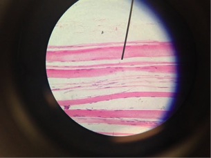 Dense Regular Connective Tissue
 Description: Parallel collagen fibers; a few elastic fibers; major cell type is the fibroblast
Function: Attaches muscle to bones or to muscles; attaches bones to bones; withstands great tensile stress when pulling force is applied in one direction
Location: Tendons, most ligaments, aponeuroses
<Marieb, Mitchell. Human Anatomy and Physiology Laboratory Manual. Pg. 79.>
Function: Attaches muscle to bones or to muscles; attaches bones to bones; withstands great tensile stress when pulling force is applied in one direction
Location: Tendons, most ligaments, aponeuroses
<Marieb, Mitchell. Human Anatomy and Physiology Laboratory Manual. Pg. 79.>
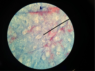
 Dense Irregular Connective Tissue
 Description: irregularly arranged collagen fibers; some elastic fibers; major cell type is the fibroblast.
Function: Able to withstand tension exerted in many directions; provides structural strength
Location: Dermis of the skin; sub mucosa of digestive tract; fibrous capsules of organs and of joints.
<Marieb, Mitchell. Human Anatomy and Physiology Laboratory Manual. Pg. 80.>
Function: Able to withstand tension exerted in many directions; provides structural strength
Location: Dermis of the skin; sub mucosa of digestive tract; fibrous capsules of organs and of joints.
<Marieb, Mitchell. Human Anatomy and Physiology Laboratory Manual. Pg. 80.>
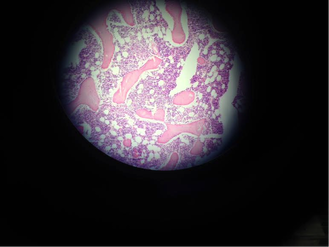
 Areolar Loose Connective Tissue
 Description: Gel-like matrix with all three fiber types; cells: (fibroblasts, macrophages, mast cells, some white blood cells) elastic fibers, collagen fibers
Function: Wraps and cushions organs; its macrophages phagocytize bacteria; plays important role in inflammation, holds and conveys tissue fluid
Location: Widely distributed under epithelia of body (forms lamina propria of mucous membranes) packages of organs; surrounds capillaries.
<Marieb, Mitchell. Human Anatomy and Physiology Laboratory Manual. Pg. 78.>
 Elastic Cartilage
 Description: Similar to hyaline cartilage but more elastic fibers in matrix.
Chondroblasts, Chondrocytes, Collagen fibers, Elastic Fibers
Function: Maintains the shape of a structure while allowing great flexibility
Location: Supports the external ear (pinna); epiglottis.
<Marieb, Mitchell. Human Anatomy and Physiology Laboratory Manual. Pg. 81.>
Chondroblasts, Chondrocytes, Collagen fibers, Elastic Fibers
Function: Maintains the shape of a structure while allowing great flexibility
Location: Supports the external ear (pinna); epiglottis.
<Marieb, Mitchell. Human Anatomy and Physiology Laboratory Manual. Pg. 81.>
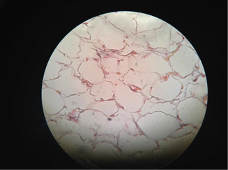 Adipose Loose Connective Tissue
 Description: Matrix as in areolar, but very sparse; closely packed adipocytes (fat cells); have nucleus pushed to the side by large fat droplet.
Function: Provides reserve fuel; insulates against heat loss; supports and protects organs
Location: Under skin; around kidneys and eyeballs; within abdomen; in breasts.
<Marieb, Mitchell. Human Anatomy and Physiology Laboratory Manual. Pg. 78.>
Function: Provides reserve fuel; insulates against heat loss; supports and protects organs
Location: Under skin; around kidneys and eyeballs; within abdomen; in breasts.
<Marieb, Mitchell. Human Anatomy and Physiology Laboratory Manual. Pg. 78.>
 Fibrocartilage
 Description: Matrix similar to but less firm than that in hyaline cartilage; thick collagen fibers predominate
Chondrocytes, Chondroblasts
Function: Tensile strength with the ability to absorb compressive shock
Location: Intervertebral discs; pubic symphysis; discs of knee joint
<Marieb, Mitchell. Human Anatomy and Physiology Laboratory Manual. Pg. 81.>
Chondrocytes, Chondroblasts
Function: Tensile strength with the ability to absorb compressive shock
Location: Intervertebral discs; pubic symphysis; discs of knee joint
<Marieb, Mitchell. Human Anatomy and Physiology Laboratory Manual. Pg. 81.>
 Reticular Loose Connective Tissue
 Description: Network of reticular fibers in a typical loose ground substance; reticular cells lie on the network
Lymphocytes, Macrophages
Function: Fibers form a soft internal skeleton (stroma) that supports other cell types including white blood cells, mast cells and macrophages.
Location: Lymphoid organs (lymph nodes, bone marrow and spleen)
<Marieb, Mitchell. Human Anatomy and Physiology Laboratory Manual. Pg. 79.>
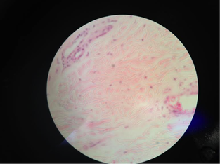
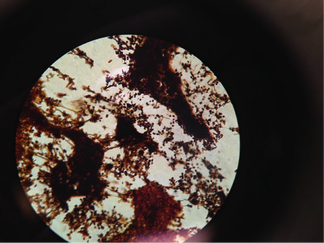
 Blood
Blood Description: Red and white blood cells in fluid matrix (plasma)
Erythrocytes, Leukocytes
Functions: Transport of respiratory gases, nutrients, wastes and other substances
Location: Contained within blood vessels
*slide might show a couple bigger cells (white blood cells) surrounded by multiple red blood cells - the filling space is plasma*
<Marieb, Mitchell. Human Anatomy and Physiology Laboratory Manual. Pg. 82.>
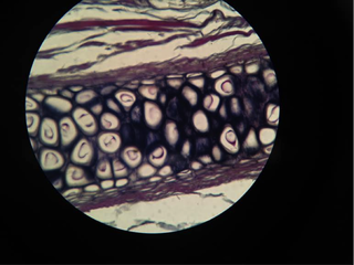
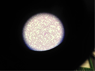
 Bone Decalcified
 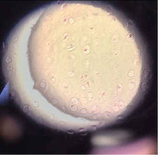 Hyaline Cartilage
 Description: Amorphous but firm matrix; collagen fibers form an imperceptible network; chondroblasts produce the matric and when mature lie in lacunae
Chondrocytes
Function: Supports and reinforces; has resilient cushioning properties; resists compressive stress
Location: Forms most of the embryonic skeleton; covers the ends of long bones in joint cavities; forms costal cartilages of the ribs; cartilages of the nose, trachea, and larynx.
*Picture is a little unclear, while studying slides look for the Chondrocyte in lacuna and the matrix*
<Marieb, Mitchell. Human Anatomy and Physiology Laboratory Manual. Pg. 80.>
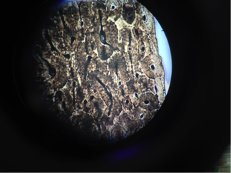 Bone (Osseous tissue)
 Description: Hard, calcified matrix containing many collagen fibers, osteocytes lie in lacunae. Very well vascularized.
Function: Bone supports and protects (by enclosing); provides levers for the muscles to act on; stores calcium and other minerals and fat; marrow inside bones is the site for blood cell formation (hematopoiesis)
Location: Bones
<Marieb, Mitchell. Human Anatomy and Physiology Laboratory Manual. Pg. 82.>


 RSS Feed