The Language of Anatomy
Anterior Terms (Front of the body)
| Axial - central part of the body (head, neck, and trunk) Appendicular - the bodies limbs and attachments on the body (arms, legs, fingers, toes) Abdominal - anterior (front) on the trunk and inferior (below) to the ribs Acromial - point of shoulder Antebrachial - your forearm Antecubital - front of the elbow while arm is extended straight Axillary - armpit Brachial - arm *test might point to the spot above your antecubital* Buccal - cheek Carpal - wrist | Cervical - neck Coxal - hip Crural - leg *this is defined as the the leg beyond the knee* Digital - fingers or toes Femoral - thigh Fibular - side of leg *also known as peroneal; test might point to the side of your calf above the ankle* Frontal - forehead Hallux - great toe Inguinal - groin *test might point to area closest to pubic* Mammary - breast Mental - chin | Nasal - nose Oral - mouth Orbital - eye Palmar - palm of the hand Patellar - kneecap (anterior knee) Pedal - foot Pelvic - pelvis region *test might point directly above pubic* Pollex - thumb Pubic - genital region *penis or vagina area* Sternal - breastbone Tarsal - ankle Thoracic - chest Umbilical - navel (belly button) |
Posterior Terms (Back of the body)
| Acromial - point of shoulder Brachial - arm *test might point to back of arm, above elbow* Calcaneal - heel of foot Cephalic - the entire back of head Dorsum - the entire back Femoral - thigh Gluteal - buttocks | Lumbar - (loin) area of your back *test might point between the ribs and hips* Manus - hand *if model is in anatomical position, test will be asking for the entire top side of hand, because it is posterior* Occipital - back of head(base of skull) Olecranal - elbow (from back view) | Otic - ear Perineal - between the anus and external genitalia Plantar - bottom of foot Popliteal - back of knee Sacral - between hips, above buttocks Scapular - shoulder blade Sural - calf Vertebral - spinal column |
Body Orientation
| Superior - (above) placement of a structure along the long axis of the body, always above other structures *on a human model, this will show the occipital is superior to the plantar; on a four-legged animal model, this will show the dorsum of an animal is superior to the sternum* Inferior - (below) along the long axis of the body, always below other structures *on a human model, the plantar is inferior to the occipital; on a four-legged animal model, the sternum is inferior to the dorsum* Anterior - (front) structures on the front of the body such as face, patellar, umbilical *on a human model, the trachea is anterior to the spine; on a four-legged animal model, the nasal is anterior to the gluteal* Posterior - (back) structures on the back of the body such as popliteal, dorsum, gluteal *on a human model, the spine is posterior to the trachea; on a four-legged animal model, the gluteal is posterior to the nasal* Medial - toward the midline *the sternum is medial to the ribs* Lateral - away from the midline *the ear is lateral to the nose* | Cephalad A. Crainal - toward the head B. Caudal - toward the tail *referring to a human, these terms are used with superior and inferior; in four-legged animals, these terms are synonymous with anterior and posterior* Dorsal - the backside of any human or animal model *on a human and animal, the word dorsal is used to describe the back* Ventral - the belly side *on a human and animal, the word ventral is used to describe the belly* *on a four-legged animal, the words dorsal and ventral are synonymous with inferior and posterior* Proximal - closer to the trunk of the body Distal - farther away from the trunk of the body *the fingers are distal to the elbow; the knee is proximal to the toes* *these terms can also be used to indicate regions (closer to or farther from the head)* Superficial - (external) toward/at the body surface Deep - (internal) away from the body surface *the skin is superficial to the skeletal muscles; the lungs are deep to the rib cage* <Marieb, Mitchell. Human Anatomy and Physiology Laboratory Manual. Pg. 4.> |
Body Planes and Sections
| The Sagittal Plane (Median, Midsagittal) Runs longitudinally and divides the body into equal left and right parts | The Transverse Plane (Cross section) Runs horizontally, divides body into superior and inferior parts *Cross sections is when organs are sectioned along this plane* | The Frontal Plane (Coronal plane) Longitudinal plane that divides the body into anterior and posterior parts |
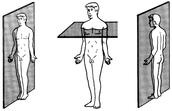
Photo from Basic Human Anatomy. Guide 1. Introduction. Web. 6 Feb. 2014.
<http://operationalmedicine.org/anatomy/Study_Guides/md0006_img_3.png>
<http://operationalmedicine.org/anatomy/Study_Guides/md0006_img_3.png>
Body Cavities
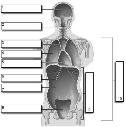
Photo from Biology Corner. Body Cavities. Web. 6 Feb. 2014.
<http://www.biologycorner.com/anatomy/intro/body_cavities_label.html>
<http://www.biologycorner.com/anatomy/intro/body_cavities_label.html>
1. Cranial Cavity - brain is enclosed by skull
2. Vertebral Cavity - (spinal cavity) spinal cord is protected by the bony vertebral column
*these two cavities are continuous with each other because the spinal cord is a continuation of the brain*
3. Mediastinum - near the median sagittal plane of chest; extends from sternum in front to vertebral column behind; contains all thoracic viscera except lungs; divided into a superior and inferior part
4. Pleural Cavity - composed of the layers of the membrane lining the lung and the chest cavity
5. Pericardial Cavity - (within the mediastinum)
6. Diaphragm - separates the chest from the abdomen; located in thoracic cavity; attached to the spine, ribs and sternum; main muscle of respiration
7. Abdominal Cavity - area surrounding the stomach, intestines, liver, and other organs
8. Pelvic Cavity - contains the reproductive organs, bladder and rectum (partially enclosed by the bony pelvis)
9. Abdominopelvic Cavity - inferior to the diaphragm
10. Ventral Body Cavity - subdivided by #3, #4, #5, #6, #7, #8, and #9
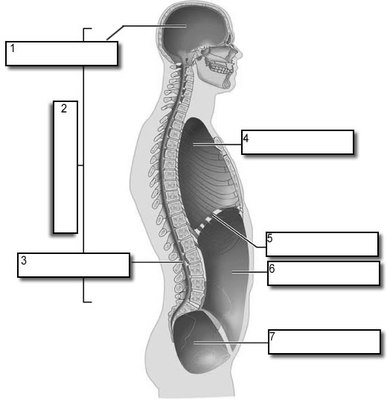
Photo from Biology Corner. Body Cavities. Web. 6 Feb. 2014.
<http://www.biologycorner.com/anatomy/intro/body_cavities_label.html>
<http://www.biologycorner.com/anatomy/intro/body_cavities_label.html>
1. Cranial Cavity
2. Dorsal Cavity
3. Vertebral Canal
4. Thoracic Cavity
5. Diaphragm
6. Abdominal Cavity
7. Pelvic Cavity
*refer to descriptions in anterior view of body cavities*
2. Dorsal Cavity
3. Vertebral Canal
4. Thoracic Cavity
5. Diaphragm
6. Abdominal Cavity
7. Pelvic Cavity
*refer to descriptions in anterior view of body cavities*
Other body cavities:
Oral Cavity - (mouth) contains the tongue and teeth, continuous with digestive tube that opens to the exterior at the anus
Nasal Cavity - (nose) part of the passages of the respiratory system
Orbital Cavity - (eye) in the skull, houses the eye
Middle Ear Cavities - each middle ear cavity lies medial to an eardrum, contain tiny bones that transmit sound vibrations to the organ of hearing in the inner ears
Synovial Cavities - in a joint between neck and vertebrae
Abdominopelvic Quadrants and Regions
Photo from The Crime Lab. Regions and Quadrants. Web. 6 Feb. 2014.
<http://shs2.westport.k12.ct.us/forensics/02-evidence/regions_&_quadrants.htm>
<http://shs2.westport.k12.ct.us/forensics/02-evidence/regions_&_quadrants.htm>
| *Left picture in order from top left, top right, bottom left, bottom right* 1. Right Upper Quadrant 2. Left Upper Quadrant 3. Right Lower Quadrant 4. Left Lower Quadrant | 1. Right Hypochondriac Region (*shown in picture as top left*) contains liver and gallbladder 2. Epigastric Region (*shown in picture as top middle*) superior to umbilical, most of stomach 3. Left Hypochondriac Region (*shown in picture as top right*) contains diaphragm 4. Right Lumbar Region (*shown in picture as middle left*) contains ascending colon of large intestine | 5. Umbilical Region (*shown in picture as middle middle*) contains transverse colon of large intestine, small intestine 6. Left Lumbar Region (*shown in picture as bottom left*) contains descending colon of large intestine 7. Right iliac (inguinal) region (*shown in picture as bottom left*) contains cecum 8. Hypogastric (pubic) region (*shown in picture as bottom middle*) contains appendix and urinary bladder 9. Left iliac (inguinal) region (*shown in picture as bottom right*) contains initial part of sigmoid colon |
Nervous Tissue
Nervous Tissue carries out the informational signaling in an organism. Our brain is made of nervous tissue, and it can choose to tell our muscles (via the spinal cord and nerves) to carry out certain actions. Our sensory systems all provide information to our brains via nervous system cells as well. (Tamarkin, Dawn)
The two types of nervous tissue are:
1. Neurons - electrically active, signaling cells of the nervous system
2. Neuroglia (Glia) - support cells;
"Nervous tissue receive nutrients from the blood. The neurons of nervous tissue are unable to divide, ever. Once they die, they cannot be replaced. Glia are able to reproduce, but are unable to take over the function of neurons. While looking at nervous tissue it looks like there are a lot of spaces where extracellular material could be present, however this is not the case. Neurons (and glia) can send out so many branches of their cells that all the space you see is full of neuronal (and glial) processes." (Tamarkin, Dawn)
Tamarkin, Dawn. STCC FOUNDATION PRESS. Web. 6 Feb. 2014. <http://faculty.stcc.edu/AandP/AP/AP1pages/Units1to4/unit3/nervous.htm>
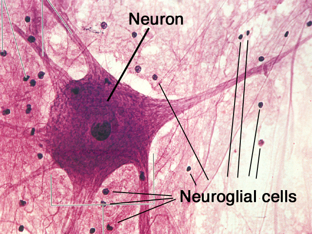
Photo from <http://www.hartnell.cc.ca.us/faculty/aedens/Bio6L/Bio6Lnervous.html>
TEST 1 REVIEW
CH 1
- Define anatomy and physiology
Anatomy is the study of the structure and relationship between body parts.
Physiology is the study of the function of body parts and the body as a whole.
- Explain the relationship between anatomy and physiology, and describe various specialties of each discipline.Anatomy:
Gross (macroscopic) anatomy is the study of body parts visible to the naked eye, such as the heart or bones.
Histology is the study of tissues at the microscopic level.
Cytology is the study of cells at the microscopic level.
Physiology:
Neurophysiology is the study of how the nervous system functions.
Cell Physiology is the study of the functions of cells at chemical and molecular levels
Systemic Physiology is the study of functions of specific organ systems
Pathological Physiology is the study of effects of diseases on organ or system functions
- Identify the major levels of organization in organisms, from the simplest to the most complex, and identify major components of each organ system.
The levels of organization of living things, from smallest to largest, are:
1. Atoms, the smallest functional units of matter.
2. Molecules, active chemicals.
3. Organelles, specialized structures within a cell.
4. Cells, the smallest living units.
5. Tissues, a group of similar cells that work together.
6. Organs, two or more tissue types working together.
7. Organ systems, two or more organs working together.
8. Organism, a single individual, including all of the above.
• Systemic Human Anatomy – will be our approach
The human body is divided into 11 interconnected organ systems. All organ
systems work together, and many organs function in more than 1 organ system.
- Explain the concept of homeostasis
The foundation of all physiology is homeostasis (“staying the same”). When the
body does not function within its normal range, organ systems malfunction,
resulting in disease.
• As our internal and external environment changes, physiological systems work
together to maintain a stable internal environment, the condition of homeostasis.
For example, systems monitor and adjust the volume and composition of body
fluids, and keep body temperature within normal limits.
• A homeostatic regulatory mechanism consists of 3 parts:
1. Receptors, sensors that respond to a stimulus.
2. The control center, receives information from sensors and sends out
commands.
3. Effectors, the cell or organ that responds to the control center.
- Describe how negative feedback and positive feedback are involved in homeostatic regulation, and explain the significance of homeostasis.
When the response of an effector opposes the original stimulus, that is called
negative feedback because it negates the stimulus.
• An example of negative feedback is the temperature thermostat in your home.
• Temperature sensors turn the air conditioner off and on to maintain air
temperature within a specific, limited range.
• In the same way, the brain controls normal body-temperature homeostasis by
negative feedback.
• When body temperature is too high or too low, the control center instructs an
effector to oppose the effects of the stimulus by increasing or decreasing blood
flow and sweat production.
• In the opposite response, positive feedback, the effector adds to the initial
stimulus instead of negating it, speeding up the process.
Positive feedback examples include, blood clotting (a little clotting provokes
more clotting) and the birthing process (contractions get closer).
• The body is constantly working, changing and responding to stimuli, a state of
dynamic equilibrium.
• All body systems must work together (systems integration) to maintain homeostasis.
- Body planes and sections are used to describe how the body or an organ is divided into two parts:
*see image of body planes*
Sagittal planes divide a body or organ vertically into right and left parts. If the right and left parts are equal, the plane is a midsagittal plane; if they're unequal, the plane is a parasagittal plane.
A frontal (coronal) plane divides the body or organ vertically into front (anterior) and rear (posterior) parts.
A horizontal (transverse) plane divides the body or organ horizontally into top (superior) and bottom (inferior) parts. This is also known as a cross‐section.
• Body cavities are enclosed areas that house organs. These cavities are organized into two groups:
• The posterior/dorsal body cavity includes the cranial cavity (which contains the brain) and the vertebral cavity (which contains the spinal cord).
• The anterior/ventral body cavity includes the thoracic cavity (which contains the lungs, each in its own pleural cavity, and the heart, in the pericardial cavity) and the abdominopelvic cavity (which contains the digestive organs in the abdominal cavity and the bladder and reproductive organs in the pelvic cavity).
• Regional terms identify specific areas of the body. In some cases, a descriptive word is used to identify the location. For example, the axial region refers to the main axis of the body—the head, neck, and trunk. The appendicular region refers to the appendages—the arms and legs. Other regional terms use a body part to identify a particular region of the body. For example, the nasal region refers to the nose.
- Identify the major body cavities and their subdivisions, and describe the functions of each.
Internal compartments called body cavities protect internal organs, hold them in
place, and allow them to change size and shape. All the internal organs found
within these cavities are called viscera.
• Moist layers of connective tissue called serous membrane cover both the walls of
internal cavities (parietal layer) and the visceral organs themselves (visceral
layer), providing a double layer of membrane between an organ and its
surroundings. Serous membrane contains a watery lubricant that reduces friction,
allowing organs to expand and contract freely.
The ventral body cavity (coelom) is divided by the diaphragm muscle into 2 parts:
1. A superior thoracic cavity, containing the
(a) pleural cavity (left and right, divided by the mediastinum)
organs: lungs
membranes: visceral and parietal pleura
(b) pericardial cavity
organs: heart
membranes: visceral and parietal pericardium
2. and an inferior abdominopelvic cavity, containing the
(a) peritoneal cavity
membranes: visceral and parietal peritoneum
(b) abdominal cavity (superior peritoneal)
organs: liver, stomach, spleen, intestine
(c) pelvic cavity (inferior peritoneal)
organs: intestine, bladder, reproductive organs.
CH 4
- Identify the four major types of tissues in the body and describe their roles
A tissue is a group of cells that have a similar shape and function. Different types of tissues can be found in different organs. In humans, there are four basic types of tissue: epithelial, connective, muscular, and nervous tissue. There may be various sub-tissues within each of the primary tissues.
Epithelial tissue covers the body surface and forms the lining for most internal cavities. The major function of epithelial tissue includes protection, secretion, absorption, and filtration. The skin is an organ made up of epithelial tissue which protects the body from dirt, dust, bacteria and other microbes that may be harmful. Cells of the epithelial tissue have different shapes as shown on the student's worksheet. Cells can be thin, flat to cubic to elongated.
Connective tissue is the most abundant and the most widely distributed of the tissues. Connective tissues perform a variety of functions including support and protection. The following tissues are found in the human body, ordinary loose connective tissue, fat tissue, dense fibrous tissue, cartilage, bone, blood, and lymph, which are all considered connective tissue.
There are three types of muscle tissue: skeletal, smooth, and cardiac. Skeletal muscle is a voluntary type of muscle tissue that is used in the contraction of skeletal parts. Smooth muscle is found in the walls of internal organs and blood vessels. It is an involuntary type. The cardiac muscle is found only in the walls of the heart and is involuntary in nature.
Nerve tissue is composed of specialized cells which not only receive stimuli but also conduct impulses to and from all parts of the body. Nerve cells or neurons are long and string-like.
In tissues the simplest combination is called a membrane, or a sheet of tissues which cover or line the body surface or divide organs into parts. Examples include the mucous membrane which lines body cavities. Tissues combine to form organs. An organ is a part of the body which performs a definite function. The final units of organization in the body are called systems. A system is a group of organs each of which contributes its share to the function of the body as a whole.
- Discuss the types and functions of epithelial tissue/Describe the relationship between form and function for each type of epithelium
Epithelial tissue covers the whole surface of the body. It is made up of cells closely packed and ranged in one or more layers. This tissue is specialised to form the covering or lining of all internal and external body surfaces. Epithelial tissue that occurs on surfaces on the interior of the body is known as endothelium. Epithelial cells are packed tightly together, with almost no intercellular spaces and only a small amount of intercellular substance. Epithelial tissue, regardless of the type, is usually separated from the underlying tissue by a thin sheet of connective tissue; basement membrane. The basement membrane provides structural support for the epithelium and also binds it to neighbouring structures.
Types of Epithelial TissueEpithelial tissue can be divided into two groups depending on the number of layers of which it is composes. Epithelial tissue which is only one cell thick is known as simple epithelium. If it is two or more cells thicksuch as the skin, it is known as stratified epithelium.
Simple epithelium
Simple epithelium can be subdivided according to the shape and function of its cells.
Functions of Epithelial Tissue
- Compare the structures and functions of the various types of connective tissues
Connective tissues vary widely in appearance and function, but they all share three basic components:
1. specialized cells
2. extracellular protein fibers
3. a fluid known as ground substance
Fibroblast most common kind of fixed cell; large, star-shaped; produces fibers by secreting protein into connective tissue matrix.
Macrophage originate as white blood cells and are almost as numerous as fibroblasts in some connective tissues; usually attached to fibers, but can detach and move about; specialized to carry on phagocytosis.
Mast Cells usually located near blood vessels; release heparin and histamine; also produce prostaglandins.
Adipocytes fat cells; store lipid droplets.
Mesenchymal Cells stem cells that are present in many connective tissues; respond to local injury or infection by dividing to produce daughter cells that differentiate into fibroblasts, macrophages, or other connective tissue cells.
Melanocytes synthesize and store the brown pigment melanin.
Lymphocytes produce antibodies; immune function.
Microphages phagocytic blood cells; when infection or injury occurs.
Collagenous Fibers thick threads of the protein collagen.
Dense Connective Tissue abundant collagenous fibers --> appear white in color.
Loose Connective Tissues sparse collagenous fibers.
Elastic Fibers bundles of microfibrils embedded in a protein called elastin.
Reticular Fibers very thin collagenous fibers; highly branched and form delicate supporting networks in tissues; found in liver and spleen which are made of pulpy tissue.
Connective Tissue Proper includes those connective tissues with many types of cells and extracellular fibers in a syrupy ground substance --> matrix.
Fluid Connective Tissues distinctive populations of cells suspended in a watery matrix that contains dissolved proteins --> blood and lymph.
Supporting Connective Tissues have a less diverse cell population and a matrix containing much more densely packed fibers; protect soft tissues and support the weight of part or all of the body --> bone or cartilage.
- Describe how cartilage and bone function as a supporting connective tissue
Bones and cartilages are types of connective tissue in the body. Bones are hard tissue that form the skeletal structure of the body; Cartilages are not as hard and rigid as bones, and present in areas of the body like the ear, nose and joints. In the joints of the body, cartilage covers the ends of the bones and acts as a shock absorber to prevent the ends of bones from rubbing against each other.
Bone
Types: Bones are either compact or spongy. Bones are classified into long, short, flat, irregular, sesamoid and sutural bones.
Functions: Protect the body against mechanical damage, assist in the movement of the body, provid a framework and shape for the body, store minerals, and produce red blood & white blood cells.
Structures: Bones are made up mostly of osteoblasts (progenitor cells), osteocytes (mature bone cells), and osteoclasts (large cells that breakdown bone tissue for growth and repair). A bone is highly vascularized.
Locations: Bones make up the majority of the axial and appendicular skeleton.
Fibrous Covering: Periostium, rich in sensory nerve endings.
Cartilage
Types: Hyaline cartilage, fibrocartilage and elastic cartilage.
Functions: Reducing friction at joints, supporting the respiratory tract, acting as shock absorbers between weight-bearing bones, and maintaining the shape and flexibility of fleshy appendages.
Structures: Cartilages comprise chondroblasts, (precursor cells), chondrocytes, and a dense matrix of collagen and elastic fibers, in which the mature chondrocytes are embedded. Cartilage is avascular.
Locations: Cartilage is a much softer, more pliable component that is mostly found in between joints of bones (articular cartilage), along the respiratory tract, and in a few other places where flexibility is needed.
Fibrous Covering: Perichondrium (but does not surround articular cartilage)
Differences in Physical structure
The structure of bones is a combination of living and dead cells embedded in a matrix. The outer hard layer of the bone is referred to as compact bone and has few spaces. The internal part of bone also called spongy tissue is porous and harbours the bone marrow and blood vessels. Other tissue found in bone includes endosteum, periosteum and nerves. Bone matrix has organic (mineral) components and inorganic components such as collagen. Bone formation is the result of hardening of this matrix.
Cartilage essentially consists of chondrocyte cells which produces extracellular matrix consisting of collagen fibres, proteoglycan and elastin fibres and different types of cartilage contain these components in different proportions. Cartilage, unlike bones, does not contain blood vessels.
Cellular Structure of Bones vs Cartilages Bone or osseous tissue is made up of osteoblasts, the progenitor cells, which give rise to osteocytes, which are the mature bone cells, and osteoclasts which are large cells that breakdown bone tissue for growth, repair and remodelling. Another type of bone lining cells is present that regulate the movement of calcium and phosphate in and out of the bone.
Cartilage comprises of chondrocytes, produced by precursor cells known as chondroblasts. Chondroblast, produce a dense matrix comprising of collagen and elastin fibres, in which the mature chondrocyte cells are embedded.
Cartilage vs Bone Diseases
Main disorders of the bone consists of are osteoporosis, in which the bone mineral density gets reduced, and increases the likelihood of a fracture, osteosarcoma, a cancerous condition of the bone, osteomyelitis, which is an infection of the bone marrow and osteogenesis imperfecta which is a genetic disorder.
Diseases of the cartilage include, osteoarthritis is a condition in which the cartilage gets thinned out resulting in friction between bones, achondroplasia, which leads to dwarfism, costochondritis, which is inflammation of the cartilage in the ribs resulting in chest pain and chondrodystrophies which are a group of diseases due to disturbance in growth of cartilage and subsequent ossification of cartilage.
Types of bones and cartilages
Differences in Function
Bones perform a variety of functions like protecting the body against mechanical damage, for example the skull protects the brain and the rib cage protects the internal organs and so on; assists in the movement of the body as the skeletal muscles are attached to the bones; provides a framework and shape of the body; stores minerals like calcium and phosphorous; red bone marrow stored in the bones, produces erythrocytes (red blood cells) and leucocytes (white blood cells); and yellow bone marrow which also contains adipose cells is an important reserved for energy.
The main functions of cartilage tissue include reducing friction at the joints, supporting tracheal and bronchial tubes, acting as shock absorbers between vertebrae, maintaining the shape and flexibility of ear, nose and so on.
Bones are classified into long, short, flat, irregular, sesamoid and sutural bones. Most bones of the limb are long bones characterized by a long shaft and curved structure. Examples include femur, tibia, fibula, humerus, ulna and radius. Short bones are the same in length and width and present in the ankle and wrist. Flat bones are thin and curved and examples include bones of the skull and sternum. Irregular bones are found in the spine and hip region. Sesamoid bones are developed in the tendons, and most commonly found in the palm of the hands and soles of the feet, and in the kneecaps. Sutural bones are very small bones found in the sutures between the cranial bones, and vary in different persons.
Types of cartilage include hyaline cartilage, fibrocartilage and elastic cartilage. Hyaline cartilage is the most abundant of the three types of cartilage. It is found mainly in the bronchial tubes, larynx, nose and trachea, end of long bones and in the embryonic skeleton. It serves to provide structure and smooth movement. Fibrocartilage is a tough form of cartilage found at the site of fractures, intervertebral discs, knee, hip and other joints. Fibrocartilage provides rigidity and structure to the attached structures. Elastic cartilage is more elastic and is present in the external ear, auditory tubes and epiglottis.
- Explain how epithelial and connective tissues combine to form four types of tissue membranes, and specify the functions of each/Describe how connective tissue establishes the framework of the body
Mucous Membranes line passageways and chambers that communicate with the exterior, including those in the digestive, respiratory, reproductive, and urinary tracts.
• Line passageways that have external connections
• In digestive, respiratory, urinary, and reproductive tracts
• Epithelial surfaces must be moist
• To reduce friction
• To facilitate absorption and excretion
• Lamina propria
• Is areolar tissue
Serous Membranes line the sealed, internal subdivisions of the ventral body cavity -- cavities not open to the exterior.
• Line cavities not open to the outside
• Are thin but strong
• Have fluid transudate to reduce friction
• Have a parietal portion covering the cavity
• Have a visceral portion (serosa) covering the organs
Pleura lines the pleural cavities and covers the lungs.
Peritoneum lines the peritoneal cavity and covers the surfaces of the enclosed organs.
• Covers abdominal organs
Pericardium lines the pericardial cavity and covers the heart.
Cutaneous Membrane skin; covers the surface of the body.
• Thick, waterproof, and dry
Synovial Membranes lines the joint cavities of bones. Produces a fluid called synovial fluid which lubricates the joint cavity.
• Line moving, articulating joint cavities
• Produce synovial fluid (lubricant)
• Protect the ends of bones
• Lack a true epithelium
- Describe the three types of muscle tissue and the special structural features of each type
Main Function of the Muscular System:
The main function of the muscular system is movement.
There are three types of muscle tissue: skeletal, cardiac, and smooth.
Skeletal Muscle-
Skeletal muscle is voluntary.
- These muscles are attached to bones and are responsible for their movement.
- Skeletal muscle consists of long fibers and is striated due to the repeating patterns of the myofilaments (made of the protein actin and myosin) that make up the fibers.
Cardiac Muscle-
Cardiac muscle is found in the heart.
- Cardiac muscle is striated like skeletal muscle, but differs in that the plasma membrane of the cardiac muscle causes the muscle to beat even when away from the heart.
- The action potentials of cardiac and skeletal muscles also differ.
Smooth Muscle-
Smooth muscle is involuntary.
- It is found in organs and enables functions such as digestion and respiration.
- Unlike skeletal and cardiac muscle, smooth muscle is not striated.
- Smooth muscle has less myosin and does not generate as much tension as skeletal muscle.
- Discuss the basic structure and role of neural tissue
Neural tissue is specialized for the conduction of electrical impulses that convey information or instructions from one region of the body to another.
About 98% of neural tissue is concentrated in the brain and spinal cord, which are the control centers for the nervous system.
Neural tissue cell types
CH 5
- Know the main structural features of the epidermis, and explain the functional significance of each
The epidermis is an avascular stratified squamous epithelium that relies on diffusion of nutrients and oxygen from capillaries in the dermis.
The most abundant cells in the epidermis are the keratinocytes (so called because they contain large amounts of the protein keratin).
Most of the body is covered by thin skin, which has only 4 layers of keratinocytes in the epidermis.
The palms of the hands and soles of the feet are covered with thick skin, which has 5 layers of keratinocytes in its epidermis.
The 5 layers or strata of keratinocytes in the thick skin of the epidermis are (from the deep basal lamia to the free surface) the:
1. stratum germinativum
2. stratum spinosum
3. stratum granulosum
4. stratum lucidum, and the
5. stratum corneum
The stratum germinativum (“germinative layer”) is attached to the basal lamina by hemidesmosomes, forming a strong bond between the epidermis and the dermis.
- Stratum germinativum forms epidermal ridges that determine our fingerprint pattern, and dermal papillae or tiny mounds. Ridges and papillae increase the area of the basal lamina, strengthening the attachment between the epidermis and dermis.
- Stratum germinativum has many germinative (stem) cells, called basal cells, which replace keratinocytes that are shed at the skin’s surface.
- Skin surfaces that have no hair have many Merkel cells in their stratum germinativum that respond to touch by releasing chemicals that trigger nervous system responses.
- Melanocytes (cells containing the pigment melanin) are also scattered throughout the stratum germinativum.
The stratum spinosum (“spiny layer”) consists of cells produced by division of the stem cells of the stratum germinosum.
- Stratum spinosum is 8 to 10 layers of keratinocytes bound together by desmosomes. The cells are shrunken so that their cytoskeletons stick out, making them appear spiny.
- Stratum spinosum contains Langerhans cells, which are active in the immune response against microorganisms and skin cancer.
- Stratum spinosum continues to divide, increasing the thickness of the epithelium.
The stratum granulosum (“grainy layer”) stops dividing and starts producing the proteins keratin (the tough, fibrous protein that makes up hair and nails) and keratohyalin (dense granules that cross-link keratin fibers).
- Once protein fibers are produced, stratum granulosum cells dehydrate and die,
creating a tightly interlocked layer of keratin fibers surrounded by keratohyalin.
Stratum lucidum (“clear layer”) is found only in thick skin, where it covers the stratum granulosum.
- Cells of stratum lucida are flat, dense and filled with keratin.
The exposed surface of the skin is stratum corneum (“horn layer”), consisting of 15 to 30 layers of keratinized cells.
- Keratinization, the formation of a layer of dead, protective cells filled with keratin, occurs on all exposed skin surfaces except the eyes.
- It takes 15 to 30 days for a cell to move from the stratum germinosum to the stratum corneum.
- The stratum corneum is water resistant, and lasts about 2 weeks before it is shed and replaced.
- Interstitial fluid lost by evaporation through the stratum corneum is called insensible (imperceptible) perspiration. Sensible perspiration is the water produced by sweat glands.
- Water loss (dehydration) through the skin is increased by damage to the stratum corneum, such as burns and blisters (insensible perspiration), or by immersion in a hypertonic solution such as seawater (osmosis). Immersion in a hypotonic solution (freshwater) causes water gain and wrinkling by osmosis.
- Know what accounts for individual difference in skin color,and discuss the response of melanocytes to sunlight exposure
- Know the interaction between sunlight and vitamin D3 production
- Know the roles of epidermal growth factor
- Know various kinds of glands in the skin, and list the secretions of those glands
- Know how the skin responds to injury and repairs itself
Websites used:
http://faculty.sdmiramar.edu/faculty/sdccd/kpetti/Bio160/LectureNotesLabManual-PDF/01-Introduction.doc.pdf
http://www.cliffsnotes.com/sciences/anatomy-and-physiology/anatomy-and-chemistry-basics/what-is-anatomy-and-physiology
http://www.msnucleus.org/membership/html/k-6/lc/humanbio/4/lchb4_3a.html
http://www.botany.uwc.ac.za/sci_ed/grade10/mammal/epithelial.htm
http://quizlet.com/19550418/ch-4-3-compare-the-structures-and-functions-of-the-various-types-of-connective-tissues-flash-cards/
http://www.diffen.com/difference/Bone_vs_Cartilage
http://quizlet.com/19550583/ch-4-4-explain-how-epithelial-and-connective-tissues-combine-to-form-four-types-of-membranes-specify-functions-flash-cards/
http://www.google.com/url?sa=t&rct=j&q=&esrc=s&source=web&cd=4&ved=0CDoQFjAD&url=http%3A%2F%2Flearning.hccs.edu%2Ffaculty%2Fa.jameossanaie%2Fbiol2401%2Flecture-powerpoints%2Fchapter-4%2Fat_download%2Ffile&ei=7LX5Uoz8Jcbq2gXDp4GQCw&usg=AFQjCNH4acnK_ZDEA142DbW3aUemv7A7iw&bvm=bv.61190604,d.b2I
http://quizlet.com/5507486/structures-locations-and-functions-of-the-three-types-of-muscular-tissue-flash-cards/
http://www.innvista.com/health/anatomy/neural-tissue/
http://faculty.sdmiramar.edu/faculty/sdccd/kpetti/Bio160/Martini7DetailLectOutlines/05-Detailed_Lect_Out_LO.doc.pdf
CH 1
- Define anatomy and physiology
Anatomy is the study of the structure and relationship between body parts.
Physiology is the study of the function of body parts and the body as a whole.
- Explain the relationship between anatomy and physiology, and describe various specialties of each discipline.Anatomy:
Gross (macroscopic) anatomy is the study of body parts visible to the naked eye, such as the heart or bones.
Histology is the study of tissues at the microscopic level.
Cytology is the study of cells at the microscopic level.
Physiology:
Neurophysiology is the study of how the nervous system functions.
Cell Physiology is the study of the functions of cells at chemical and molecular levels
Systemic Physiology is the study of functions of specific organ systems
Pathological Physiology is the study of effects of diseases on organ or system functions
- Identify the major levels of organization in organisms, from the simplest to the most complex, and identify major components of each organ system.
The levels of organization of living things, from smallest to largest, are:
1. Atoms, the smallest functional units of matter.
2. Molecules, active chemicals.
3. Organelles, specialized structures within a cell.
4. Cells, the smallest living units.
5. Tissues, a group of similar cells that work together.
6. Organs, two or more tissue types working together.
7. Organ systems, two or more organs working together.
8. Organism, a single individual, including all of the above.
• Systemic Human Anatomy – will be our approach
The human body is divided into 11 interconnected organ systems. All organ
systems work together, and many organs function in more than 1 organ system.
- Explain the concept of homeostasis
The foundation of all physiology is homeostasis (“staying the same”). When the
body does not function within its normal range, organ systems malfunction,
resulting in disease.
• As our internal and external environment changes, physiological systems work
together to maintain a stable internal environment, the condition of homeostasis.
For example, systems monitor and adjust the volume and composition of body
fluids, and keep body temperature within normal limits.
• A homeostatic regulatory mechanism consists of 3 parts:
1. Receptors, sensors that respond to a stimulus.
2. The control center, receives information from sensors and sends out
commands.
3. Effectors, the cell or organ that responds to the control center.
- Describe how negative feedback and positive feedback are involved in homeostatic regulation, and explain the significance of homeostasis.
When the response of an effector opposes the original stimulus, that is called
negative feedback because it negates the stimulus.
• An example of negative feedback is the temperature thermostat in your home.
• Temperature sensors turn the air conditioner off and on to maintain air
temperature within a specific, limited range.
• In the same way, the brain controls normal body-temperature homeostasis by
negative feedback.
• When body temperature is too high or too low, the control center instructs an
effector to oppose the effects of the stimulus by increasing or decreasing blood
flow and sweat production.
• In the opposite response, positive feedback, the effector adds to the initial
stimulus instead of negating it, speeding up the process.
Positive feedback examples include, blood clotting (a little clotting provokes
more clotting) and the birthing process (contractions get closer).
• The body is constantly working, changing and responding to stimuli, a state of
dynamic equilibrium.
• All body systems must work together (systems integration) to maintain homeostasis.
- Body planes and sections are used to describe how the body or an organ is divided into two parts:
*see image of body planes*
Sagittal planes divide a body or organ vertically into right and left parts. If the right and left parts are equal, the plane is a midsagittal plane; if they're unequal, the plane is a parasagittal plane.
A frontal (coronal) plane divides the body or organ vertically into front (anterior) and rear (posterior) parts.
A horizontal (transverse) plane divides the body or organ horizontally into top (superior) and bottom (inferior) parts. This is also known as a cross‐section.
• Body cavities are enclosed areas that house organs. These cavities are organized into two groups:
• The posterior/dorsal body cavity includes the cranial cavity (which contains the brain) and the vertebral cavity (which contains the spinal cord).
• The anterior/ventral body cavity includes the thoracic cavity (which contains the lungs, each in its own pleural cavity, and the heart, in the pericardial cavity) and the abdominopelvic cavity (which contains the digestive organs in the abdominal cavity and the bladder and reproductive organs in the pelvic cavity).
• Regional terms identify specific areas of the body. In some cases, a descriptive word is used to identify the location. For example, the axial region refers to the main axis of the body—the head, neck, and trunk. The appendicular region refers to the appendages—the arms and legs. Other regional terms use a body part to identify a particular region of the body. For example, the nasal region refers to the nose.
- Identify the major body cavities and their subdivisions, and describe the functions of each.
Internal compartments called body cavities protect internal organs, hold them in
place, and allow them to change size and shape. All the internal organs found
within these cavities are called viscera.
• Moist layers of connective tissue called serous membrane cover both the walls of
internal cavities (parietal layer) and the visceral organs themselves (visceral
layer), providing a double layer of membrane between an organ and its
surroundings. Serous membrane contains a watery lubricant that reduces friction,
allowing organs to expand and contract freely.
The ventral body cavity (coelom) is divided by the diaphragm muscle into 2 parts:
1. A superior thoracic cavity, containing the
(a) pleural cavity (left and right, divided by the mediastinum)
organs: lungs
membranes: visceral and parietal pleura
(b) pericardial cavity
organs: heart
membranes: visceral and parietal pericardium
2. and an inferior abdominopelvic cavity, containing the
(a) peritoneal cavity
membranes: visceral and parietal peritoneum
(b) abdominal cavity (superior peritoneal)
organs: liver, stomach, spleen, intestine
(c) pelvic cavity (inferior peritoneal)
organs: intestine, bladder, reproductive organs.
CH 4
- Identify the four major types of tissues in the body and describe their roles
A tissue is a group of cells that have a similar shape and function. Different types of tissues can be found in different organs. In humans, there are four basic types of tissue: epithelial, connective, muscular, and nervous tissue. There may be various sub-tissues within each of the primary tissues.
Epithelial tissue covers the body surface and forms the lining for most internal cavities. The major function of epithelial tissue includes protection, secretion, absorption, and filtration. The skin is an organ made up of epithelial tissue which protects the body from dirt, dust, bacteria and other microbes that may be harmful. Cells of the epithelial tissue have different shapes as shown on the student's worksheet. Cells can be thin, flat to cubic to elongated.
Connective tissue is the most abundant and the most widely distributed of the tissues. Connective tissues perform a variety of functions including support and protection. The following tissues are found in the human body, ordinary loose connective tissue, fat tissue, dense fibrous tissue, cartilage, bone, blood, and lymph, which are all considered connective tissue.
There are three types of muscle tissue: skeletal, smooth, and cardiac. Skeletal muscle is a voluntary type of muscle tissue that is used in the contraction of skeletal parts. Smooth muscle is found in the walls of internal organs and blood vessels. It is an involuntary type. The cardiac muscle is found only in the walls of the heart and is involuntary in nature.
Nerve tissue is composed of specialized cells which not only receive stimuli but also conduct impulses to and from all parts of the body. Nerve cells or neurons are long and string-like.
In tissues the simplest combination is called a membrane, or a sheet of tissues which cover or line the body surface or divide organs into parts. Examples include the mucous membrane which lines body cavities. Tissues combine to form organs. An organ is a part of the body which performs a definite function. The final units of organization in the body are called systems. A system is a group of organs each of which contributes its share to the function of the body as a whole.
- Discuss the types and functions of epithelial tissue/Describe the relationship between form and function for each type of epithelium
Epithelial tissue covers the whole surface of the body. It is made up of cells closely packed and ranged in one or more layers. This tissue is specialised to form the covering or lining of all internal and external body surfaces. Epithelial tissue that occurs on surfaces on the interior of the body is known as endothelium. Epithelial cells are packed tightly together, with almost no intercellular spaces and only a small amount of intercellular substance. Epithelial tissue, regardless of the type, is usually separated from the underlying tissue by a thin sheet of connective tissue; basement membrane. The basement membrane provides structural support for the epithelium and also binds it to neighbouring structures.
Types of Epithelial TissueEpithelial tissue can be divided into two groups depending on the number of layers of which it is composes. Epithelial tissue which is only one cell thick is known as simple epithelium. If it is two or more cells thicksuch as the skin, it is known as stratified epithelium.
Simple epithelium
Simple epithelium can be subdivided according to the shape and function of its cells.
- Squamous (pavement) epithelium.Squamous cells have the appearance of thin, flat plates. The shape of the nucleus usually corresponds to the cell form and help to identify the type of epithelium. Squamous cells, for example, tend to havehorizontall flattened, elliptical nuclei because of the thin flattened form of the cell. They form the lining of cavities such as the mouth, blood vessels, heart and lungs and make up the outer layers of the skin.
- Simple Cuboidal Epithelium As their name implies, cuboidal cells are roughly square or cuboidal in shape. Each cell has a spherical nucleus in the centre. Cuboidal epithelium is found in glands and in the lining of the kidney tubules as well as in the ducts of the glands. They also constitute the germinal epithelium which produces the egg cells in the female ovary and the sperm cells in the male testes.
- Simple Columnar Epithelium Columnar epithelial cells occur in one or more layers. The cells are elongated and column-shaped. The nuclei are elongated and are usually located near the base of the cells. Columnar epithelium forms the lining of the stomach and intestines. Some columnar cells are specialised for sensory reception such as in the nose, ears and the taste buds of the tongue. Goblet cells (unicellular glands) are found between thecolumnar epithelial cells of the duodenum. They secrete mucus or slime, a lubricating substance which keeps the surface smooth.
- Ciliated Columnar Epithelium These are simple columnar epithelial cells, but in addition, they posses fine hair-like outgrowths, cilia on their free surfaces. These cilia are capable of rapid, rhythmic, wavelike beatings in a certain direction. This movement of the cilia in a certain direction causes the mucus, which is secreted by the goblet cells, to move (flow or stream) in that direction. Ciliated epithelium is usually found in the air passages like the nose. It is also found in the uterus and Fallopian tubes of females. The movement of the cilia propel the ovum to the uterus.
- Glandular Epithelium Columnar epithelium with goblet cells is called glandular epithelium. Some parts of the glandular epithelium consist of such a large number of goblet cells that there are only a few normal epithelial cells left. Columnar and cuboidal epithelial cells often become specialised as gland cells which are capable of synthesising and secreting certain substances such as enzymes, hormones, milk, mucus, sweat, wax and saliva. Unicellular glands consist of single, isolated glandular cells such as the goblet cells. Sometimes a portion of the epithelial tissue becomes invaginated and a multicellular gland is formed. Multicellular glands are composed of clusters of cells. Most glands are multicellular including the the salivary glands.
- Stratified Epithelium Where body linings have to withstand wear and tear, the epithelia are composed of several layers of cells and are then called compound or stratified epithelium. The top cells are flat and scaly and it may or may not be keratinised (i.e. containing a tough, resistant protein called keratin). The mammalian skin is an example of dry, keratinised, stratified epithelium. The lining of the mouth cavity is an example of an unkeratinisied, stratified epithelium.
Functions of Epithelial Tissue
- Protection Epithelial cells from the skin protect underlying tissue from mechanical injury, harmful chemicals, invading bacteria and from excessive loss of water.
- Sensation Sensory stimuli penetrate specialised epithelial cells. Specialised epithelial tissue containing sensory nerve endings is found in the skin, eyes, ears, nose and on the tongue.
- Secretion In glands, epithelial tissue is specialised to secrete specific chemical substances such as enzymes, hormones and lubricating fluids.
- Absorption Certain epithelial cells lining the small intestine absorb nutrients from the digestion of food.
- Excretion Epithelial tissues in the kidney excrete waste products from the body and reabsorb needed materials from the urine. Sweat is also excreted from the body by epithelial cells in the sweat glands.
- Diffusion Simple epithelium promotes the diffusion of gases, liquids and nutrients. Because they form such a thin lining, they are ideal for the diffusion of gases (eg. walls of capillaries and lungs).
- Cleaning Ciliated epithelium assists in removing dust particles and foreign bodies which have entered the air passages.
- Reduces Friction The smooth, tightly-interlocking, epithelial cells that line the entire circulatory system reduce friction between the blood and the walls of the blood vessels.
- Compare the structures and functions of the various types of connective tissues
Connective tissues vary widely in appearance and function, but they all share three basic components:
1. specialized cells
2. extracellular protein fibers
3. a fluid known as ground substance
Fibroblast most common kind of fixed cell; large, star-shaped; produces fibers by secreting protein into connective tissue matrix.
Macrophage originate as white blood cells and are almost as numerous as fibroblasts in some connective tissues; usually attached to fibers, but can detach and move about; specialized to carry on phagocytosis.
Mast Cells usually located near blood vessels; release heparin and histamine; also produce prostaglandins.
Adipocytes fat cells; store lipid droplets.
Mesenchymal Cells stem cells that are present in many connective tissues; respond to local injury or infection by dividing to produce daughter cells that differentiate into fibroblasts, macrophages, or other connective tissue cells.
Melanocytes synthesize and store the brown pigment melanin.
Lymphocytes produce antibodies; immune function.
Microphages phagocytic blood cells; when infection or injury occurs.
Collagenous Fibers thick threads of the protein collagen.
Dense Connective Tissue abundant collagenous fibers --> appear white in color.
Loose Connective Tissues sparse collagenous fibers.
Elastic Fibers bundles of microfibrils embedded in a protein called elastin.
Reticular Fibers very thin collagenous fibers; highly branched and form delicate supporting networks in tissues; found in liver and spleen which are made of pulpy tissue.
Connective Tissue Proper includes those connective tissues with many types of cells and extracellular fibers in a syrupy ground substance --> matrix.
Fluid Connective Tissues distinctive populations of cells suspended in a watery matrix that contains dissolved proteins --> blood and lymph.
Supporting Connective Tissues have a less diverse cell population and a matrix containing much more densely packed fibers; protect soft tissues and support the weight of part or all of the body --> bone or cartilage.
- Describe how cartilage and bone function as a supporting connective tissue
Bones and cartilages are types of connective tissue in the body. Bones are hard tissue that form the skeletal structure of the body; Cartilages are not as hard and rigid as bones, and present in areas of the body like the ear, nose and joints. In the joints of the body, cartilage covers the ends of the bones and acts as a shock absorber to prevent the ends of bones from rubbing against each other.
Bone
Types: Bones are either compact or spongy. Bones are classified into long, short, flat, irregular, sesamoid and sutural bones.
Functions: Protect the body against mechanical damage, assist in the movement of the body, provid a framework and shape for the body, store minerals, and produce red blood & white blood cells.
Structures: Bones are made up mostly of osteoblasts (progenitor cells), osteocytes (mature bone cells), and osteoclasts (large cells that breakdown bone tissue for growth and repair). A bone is highly vascularized.
Locations: Bones make up the majority of the axial and appendicular skeleton.
Fibrous Covering: Periostium, rich in sensory nerve endings.
Cartilage
Types: Hyaline cartilage, fibrocartilage and elastic cartilage.
Functions: Reducing friction at joints, supporting the respiratory tract, acting as shock absorbers between weight-bearing bones, and maintaining the shape and flexibility of fleshy appendages.
Structures: Cartilages comprise chondroblasts, (precursor cells), chondrocytes, and a dense matrix of collagen and elastic fibers, in which the mature chondrocytes are embedded. Cartilage is avascular.
Locations: Cartilage is a much softer, more pliable component that is mostly found in between joints of bones (articular cartilage), along the respiratory tract, and in a few other places where flexibility is needed.
Fibrous Covering: Perichondrium (but does not surround articular cartilage)
Differences in Physical structure
The structure of bones is a combination of living and dead cells embedded in a matrix. The outer hard layer of the bone is referred to as compact bone and has few spaces. The internal part of bone also called spongy tissue is porous and harbours the bone marrow and blood vessels. Other tissue found in bone includes endosteum, periosteum and nerves. Bone matrix has organic (mineral) components and inorganic components such as collagen. Bone formation is the result of hardening of this matrix.
Cartilage essentially consists of chondrocyte cells which produces extracellular matrix consisting of collagen fibres, proteoglycan and elastin fibres and different types of cartilage contain these components in different proportions. Cartilage, unlike bones, does not contain blood vessels.
Cellular Structure of Bones vs Cartilages Bone or osseous tissue is made up of osteoblasts, the progenitor cells, which give rise to osteocytes, which are the mature bone cells, and osteoclasts which are large cells that breakdown bone tissue for growth, repair and remodelling. Another type of bone lining cells is present that regulate the movement of calcium and phosphate in and out of the bone.
Cartilage comprises of chondrocytes, produced by precursor cells known as chondroblasts. Chondroblast, produce a dense matrix comprising of collagen and elastin fibres, in which the mature chondrocyte cells are embedded.
Cartilage vs Bone Diseases
Main disorders of the bone consists of are osteoporosis, in which the bone mineral density gets reduced, and increases the likelihood of a fracture, osteosarcoma, a cancerous condition of the bone, osteomyelitis, which is an infection of the bone marrow and osteogenesis imperfecta which is a genetic disorder.
Diseases of the cartilage include, osteoarthritis is a condition in which the cartilage gets thinned out resulting in friction between bones, achondroplasia, which leads to dwarfism, costochondritis, which is inflammation of the cartilage in the ribs resulting in chest pain and chondrodystrophies which are a group of diseases due to disturbance in growth of cartilage and subsequent ossification of cartilage.
Types of bones and cartilages
Differences in Function
Bones perform a variety of functions like protecting the body against mechanical damage, for example the skull protects the brain and the rib cage protects the internal organs and so on; assists in the movement of the body as the skeletal muscles are attached to the bones; provides a framework and shape of the body; stores minerals like calcium and phosphorous; red bone marrow stored in the bones, produces erythrocytes (red blood cells) and leucocytes (white blood cells); and yellow bone marrow which also contains adipose cells is an important reserved for energy.
The main functions of cartilage tissue include reducing friction at the joints, supporting tracheal and bronchial tubes, acting as shock absorbers between vertebrae, maintaining the shape and flexibility of ear, nose and so on.
Bones are classified into long, short, flat, irregular, sesamoid and sutural bones. Most bones of the limb are long bones characterized by a long shaft and curved structure. Examples include femur, tibia, fibula, humerus, ulna and radius. Short bones are the same in length and width and present in the ankle and wrist. Flat bones are thin and curved and examples include bones of the skull and sternum. Irregular bones are found in the spine and hip region. Sesamoid bones are developed in the tendons, and most commonly found in the palm of the hands and soles of the feet, and in the kneecaps. Sutural bones are very small bones found in the sutures between the cranial bones, and vary in different persons.
Types of cartilage include hyaline cartilage, fibrocartilage and elastic cartilage. Hyaline cartilage is the most abundant of the three types of cartilage. It is found mainly in the bronchial tubes, larynx, nose and trachea, end of long bones and in the embryonic skeleton. It serves to provide structure and smooth movement. Fibrocartilage is a tough form of cartilage found at the site of fractures, intervertebral discs, knee, hip and other joints. Fibrocartilage provides rigidity and structure to the attached structures. Elastic cartilage is more elastic and is present in the external ear, auditory tubes and epiglottis.
- Explain how epithelial and connective tissues combine to form four types of tissue membranes, and specify the functions of each/Describe how connective tissue establishes the framework of the body
Mucous Membranes line passageways and chambers that communicate with the exterior, including those in the digestive, respiratory, reproductive, and urinary tracts.
• Line passageways that have external connections
• In digestive, respiratory, urinary, and reproductive tracts
• Epithelial surfaces must be moist
• To reduce friction
• To facilitate absorption and excretion
• Lamina propria
• Is areolar tissue
Serous Membranes line the sealed, internal subdivisions of the ventral body cavity -- cavities not open to the exterior.
• Line cavities not open to the outside
• Are thin but strong
• Have fluid transudate to reduce friction
• Have a parietal portion covering the cavity
• Have a visceral portion (serosa) covering the organs
Pleura lines the pleural cavities and covers the lungs.
Peritoneum lines the peritoneal cavity and covers the surfaces of the enclosed organs.
• Covers abdominal organs
Pericardium lines the pericardial cavity and covers the heart.
Cutaneous Membrane skin; covers the surface of the body.
• Thick, waterproof, and dry
Synovial Membranes lines the joint cavities of bones. Produces a fluid called synovial fluid which lubricates the joint cavity.
• Line moving, articulating joint cavities
• Produce synovial fluid (lubricant)
• Protect the ends of bones
• Lack a true epithelium
- Describe the three types of muscle tissue and the special structural features of each type
Main Function of the Muscular System:
The main function of the muscular system is movement.
There are three types of muscle tissue: skeletal, cardiac, and smooth.
Skeletal Muscle-
Skeletal muscle is voluntary.
- These muscles are attached to bones and are responsible for their movement.
- Skeletal muscle consists of long fibers and is striated due to the repeating patterns of the myofilaments (made of the protein actin and myosin) that make up the fibers.
Cardiac Muscle-
Cardiac muscle is found in the heart.
- Cardiac muscle is striated like skeletal muscle, but differs in that the plasma membrane of the cardiac muscle causes the muscle to beat even when away from the heart.
- The action potentials of cardiac and skeletal muscles also differ.
Smooth Muscle-
Smooth muscle is involuntary.
- It is found in organs and enables functions such as digestion and respiration.
- Unlike skeletal and cardiac muscle, smooth muscle is not striated.
- Smooth muscle has less myosin and does not generate as much tension as skeletal muscle.
- Discuss the basic structure and role of neural tissue
Neural tissue is specialized for the conduction of electrical impulses that convey information or instructions from one region of the body to another.
About 98% of neural tissue is concentrated in the brain and spinal cord, which are the control centers for the nervous system.
Neural tissue cell types
- Neurons transmit signals as electrical charges which affect their cell membranes. A neuron has a cell body (soma) that contains a nucleus. The stimulus that results in the production of an electrical impulse usually affects the cell membrane of one of the dendrites, which then eventually travels along the length of an axon, which can be a meter long. Axons are often called nerve fibers with each ending at a synaptic terminal.
- Neuroglia are cells of the CNS (central nervous system) and PNS (peripheral nervous system) that support and protect the neurons. They provide the physical support for neural tissue, as well as maintaining the chemical composition of the tissue fluids and defending the tissue from infection.
- Neuron (nerve cell)
- cell body – contains the nucleus – regulates the functioning of the neuron
- axon – cellular process (extension) – carries impulses away from the cell body
- dendrites – cellular process (extension) – carries impulses toward the cell body
- Synapse – space between axon of one neuron and the dendrite or cell body of the next neuron – transmits impulses from one neuron to the others
- Neurotransmitters – chemicals released by axons – transmit impulses across synapses
- Neuroglia – specialized cells in the CNS – forms myelin sheaths and other functions
- Schwann cells – specialized PNS cells – forms the myelin sheaths around neurons
CH 5
- Know the main structural features of the epidermis, and explain the functional significance of each
The epidermis is an avascular stratified squamous epithelium that relies on diffusion of nutrients and oxygen from capillaries in the dermis.
The most abundant cells in the epidermis are the keratinocytes (so called because they contain large amounts of the protein keratin).
Most of the body is covered by thin skin, which has only 4 layers of keratinocytes in the epidermis.
The palms of the hands and soles of the feet are covered with thick skin, which has 5 layers of keratinocytes in its epidermis.
The 5 layers or strata of keratinocytes in the thick skin of the epidermis are (from the deep basal lamia to the free surface) the:
1. stratum germinativum
2. stratum spinosum
3. stratum granulosum
4. stratum lucidum, and the
5. stratum corneum
The stratum germinativum (“germinative layer”) is attached to the basal lamina by hemidesmosomes, forming a strong bond between the epidermis and the dermis.
- Stratum germinativum forms epidermal ridges that determine our fingerprint pattern, and dermal papillae or tiny mounds. Ridges and papillae increase the area of the basal lamina, strengthening the attachment between the epidermis and dermis.
- Stratum germinativum has many germinative (stem) cells, called basal cells, which replace keratinocytes that are shed at the skin’s surface.
- Skin surfaces that have no hair have many Merkel cells in their stratum germinativum that respond to touch by releasing chemicals that trigger nervous system responses.
- Melanocytes (cells containing the pigment melanin) are also scattered throughout the stratum germinativum.
The stratum spinosum (“spiny layer”) consists of cells produced by division of the stem cells of the stratum germinosum.
- Stratum spinosum is 8 to 10 layers of keratinocytes bound together by desmosomes. The cells are shrunken so that their cytoskeletons stick out, making them appear spiny.
- Stratum spinosum contains Langerhans cells, which are active in the immune response against microorganisms and skin cancer.
- Stratum spinosum continues to divide, increasing the thickness of the epithelium.
The stratum granulosum (“grainy layer”) stops dividing and starts producing the proteins keratin (the tough, fibrous protein that makes up hair and nails) and keratohyalin (dense granules that cross-link keratin fibers).
- Once protein fibers are produced, stratum granulosum cells dehydrate and die,
creating a tightly interlocked layer of keratin fibers surrounded by keratohyalin.
Stratum lucidum (“clear layer”) is found only in thick skin, where it covers the stratum granulosum.
- Cells of stratum lucida are flat, dense and filled with keratin.
The exposed surface of the skin is stratum corneum (“horn layer”), consisting of 15 to 30 layers of keratinized cells.
- Keratinization, the formation of a layer of dead, protective cells filled with keratin, occurs on all exposed skin surfaces except the eyes.
- It takes 15 to 30 days for a cell to move from the stratum germinosum to the stratum corneum.
- The stratum corneum is water resistant, and lasts about 2 weeks before it is shed and replaced.
- Interstitial fluid lost by evaporation through the stratum corneum is called insensible (imperceptible) perspiration. Sensible perspiration is the water produced by sweat glands.
- Water loss (dehydration) through the skin is increased by damage to the stratum corneum, such as burns and blisters (insensible perspiration), or by immersion in a hypertonic solution such as seawater (osmosis). Immersion in a hypotonic solution (freshwater) causes water gain and wrinkling by osmosis.
- Know what accounts for individual difference in skin color,and discuss the response of melanocytes to sunlight exposure
- Know the interaction between sunlight and vitamin D3 production
- Know the roles of epidermal growth factor
- Know various kinds of glands in the skin, and list the secretions of those glands
- Know how the skin responds to injury and repairs itself
Websites used:
http://faculty.sdmiramar.edu/faculty/sdccd/kpetti/Bio160/LectureNotesLabManual-PDF/01-Introduction.doc.pdf
http://www.cliffsnotes.com/sciences/anatomy-and-physiology/anatomy-and-chemistry-basics/what-is-anatomy-and-physiology
http://www.msnucleus.org/membership/html/k-6/lc/humanbio/4/lchb4_3a.html
http://www.botany.uwc.ac.za/sci_ed/grade10/mammal/epithelial.htm
http://quizlet.com/19550418/ch-4-3-compare-the-structures-and-functions-of-the-various-types-of-connective-tissues-flash-cards/
http://www.diffen.com/difference/Bone_vs_Cartilage
http://quizlet.com/19550583/ch-4-4-explain-how-epithelial-and-connective-tissues-combine-to-form-four-types-of-membranes-specify-functions-flash-cards/
http://www.google.com/url?sa=t&rct=j&q=&esrc=s&source=web&cd=4&ved=0CDoQFjAD&url=http%3A%2F%2Flearning.hccs.edu%2Ffaculty%2Fa.jameossanaie%2Fbiol2401%2Flecture-powerpoints%2Fchapter-4%2Fat_download%2Ffile&ei=7LX5Uoz8Jcbq2gXDp4GQCw&usg=AFQjCNH4acnK_ZDEA142DbW3aUemv7A7iw&bvm=bv.61190604,d.b2I
http://quizlet.com/5507486/structures-locations-and-functions-of-the-three-types-of-muscular-tissue-flash-cards/
http://www.innvista.com/health/anatomy/neural-tissue/
http://faculty.sdmiramar.edu/faculty/sdccd/kpetti/Bio160/Martini7DetailLectOutlines/05-Detailed_Lect_Out_LO.doc.pdf


 RSS Feed